Written by a CuraCore Veterinary Medical Acupuncture course graduate. Signed release obtained from client/author. 4S2019021
Abstract
Beau, a 10 year old, male neutered, mixed breed dog was presented to the Neurology service on May 11th, 2019, for further treatment of degenerative myelopathy. Beau was previously examined in February 2019 for a chronic, progressive thoracolumbar myelopathy. A magnetic resonance imaging (MRI) scan of his complete spine and genetic testing for degenerative myelopathy (DM) was performed. Beau was diagnosed with degenerative myelopathy and a formal treatment regimen of aggressive physical rehabilitation and acupuncture was recommended. Treatment was initiated with biweekly land and water treadmill sessions and weekly/biweekly acupuncture. The treatment regimen was well tolerated and Beau remains ambulatory with slowly progressive paraparesis.
History and Presentation
Beau, a 10 year old, male neutered, mixed breed dog was presented to the Neurology service on May 11th, 2019, for further treatment of suspected degenerative myelopathy (DM). In January 2019, Beau’s owner noted that he began to exhibit signs of mild hind limb weakness characterized by slipping out occasionally on hardwood floors and scuffing his pelvic limb nails on the pavement and hardwood floors when
ambulating. This weakness slowly progressed and he was initially evaluated by the Blue-Pearl Neurology service in February 2019. He was diagnosed with a myelopathy affecting the spinal segment starting at the 3rd thoracic vertebra and extending to the 3rd lumbar vertebra (T3-L3). A complete MRI of the cervical, thoracic and lumbar spine was performed. This scan was unremarkable with no extradural or intradural lesion or spinal compression noted. A genetic test for the superoxide dismutase 1 (SOD1) mutation, the mutation that causes the majority of cases of degenerative myelopathy in dogs, was performed. Beau was diagnosed as affected (carries two mutated copies) for this mutation. In previous history, Beau was diagnosed with suspected idiopathic epilepsy in 2017 after an onset of focal seizure activity. A brain MRI was performed and was normal. He has been treated with levetiracetam; 1000mg extended release, every 12 hours since that time and his current seizure frequency is well controlled. Beau has been an otherwise healthy dog with no other previous concerns noted by his owner. He lives in a two level, ranch style home with one other canine housemate. They have access to a small, fenced back yard and their exercise consists of controlled leash walks of 1-2 miles, 3-4 times weekly. Beau eats a maintenance diet of Purina Pro Plan.
Physical and Clinical Assessment
Beau was bright, alert and responsive (BAR) on presentation. His temperature, pulse, respiration, cardiac and thoracic auscultation and pulse palpation were within normal limits. His weight was 76.1 pounds. Beau’s neurological examination revealed a normal, alert mentation with a normal cranial nerve examination. Gait evaluation revealed a mild, ambulatory paraparesis with mild proprioceptive ataxia in the pelvic limbs. His thoracic limb gait was normal. Spinal reflexes including withdrawal reflexes in all four limbs and patellar reflexes were normal. Conscious proprioception was normal in the thoracic limbs and delayed in the pelvic limbs. Mild nail wear was present on the middle digits of the pelvic limbs. No evidence of pain or discomfort was noted on joint palpation. Myofascial palpation of the face and head was normal. Taut banding was
noted in the cervical musculature and upper trapezius muscles bilaterally. Palpation of the back and pelvic limbs revealed taut banding in the vastus lateralis muscles bilaterally and tenderness to palpation was noted bilaterally in the longissimus and iliocostalis muscles from the region of T12-L3. No other abnormalities were noted on physical, orthopedic, neurological and myofascial examinations.
Problem List
T3-L3 myelopathy
Focal seizures
Multifocal muscle taut banding and pain on palpation
Differential Diagnoses
1. T3-L3 myelopathy 2. Focal seizures
V – Fibrocartilagenous embolism (FCE) V – Forebrain embolism
I – Spinal empyema I – Cryptococcus infection
N – Spinal tumor N – Meningioma
D – Degenerative myelopathy D – Lafora Disease
1 – Owner medication exposure I – Flea/tick medication reaction
C – Syringomyelia C – Hydrocephalus
A – Autoimmune myelitis (meningomyelitis) A – Autoimmune encephalitis
T – Spinal fracture T – Head trauma
E – Hypothyroidism E – Hypoglycemia
M – Taut banding in the longissimus and M – Facial muscle pain leading to
iliocostalis muscles leading to back pain facial muscle twitching
Putative/Definitive Diagnoses
It was determined that Beau’s thoracolumbar myelopathy was most likely caused by the degenerative condition, degenerative myelopathy. Numerous diagnoses were ruled out using a combination of his clinical history (no history of trauma or medication exposure), blood work results including thyroid hormone evaluation (normal) and physical examination (myofascial examination did not reveal findings that would cause current neu-rologic dysfunction signs). The complete spinal MRI was also unremarkable therefore ruling out the following possible causes: intervertebral disc herniation, FCE, spi-nal infection, spinal tumor, syringomyelia, trauma and spinal fracture. Beau’s genetic test-ing revealed that he was homozygous for the SOD1 mutation. This mutation causes the majority of cases of DM in dogs. A spinal cord biopsy would be required for a
histopathological diagnosis of this disease, however alternative diagnoses would be unlikely, given the previously mentioned testing results.
The most likely definitive diagnosis for Beau’s focal seizure activity is idiopathic epilepsy. At the time of the onset of his seizure activity, blood work was normal, thereby ruling out numerous metabolic and endocrine causes. Intoxication would be unlikely as the seizure activity continued for months without history of repeated toxin exposure. Beau had a brain MRI performed which was normal with no evidence of a pre-vious vascular insult, congenital malformation, tumor, trauma, inflammation or infection.
Medical Decision Making
It was decided to focus treatment on Beau’s thoracolumbar myelopathy with limited additional parasympathomimetic points for support of his epilepsy, as this was currently well controlled and his temperament would only allow for focused treatments of limited time frame. We would address spinal cord function with both dry needling and electroacupuncture along the Bladder line of the thoracolumbar spine. We would treat his tight cervical musculature with dry needling of Bladder points in the cervical area and associated trigger points and dry needling of trigger points in the vastus lateralis muscles bilaterally. If Beau were tolerating treatments well, Bafeng points would be added bilaterally to help increase neurologic stimulation to the toes and improve proprioception. Each acupuncture treatment would be followed by massage focusing on the cervical muscles, back and pelvic limb musculature. Phototherapy of the thoracolumbar spine was recommended and Beau’s owner performed this at home on a weekly basis, as she is a certified canine rehabilitation practitioner (CCRP).
Medical Acupuncture and Related Techniques
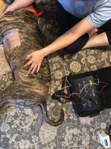
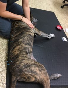
Beau was started on a weekly/biweekly treatment regimen to include dry needling, electroacupuncture and massage. This schedule was chosen to accommodate both the recommendation for consistent treatment and his owner’s schedule. He was treated with the following points: GV 20, GV 14, Bai Hui, bilateral BL 10, BL 18, BL 20, BL 23, BL25, BL 27, BL 28, ST 36 (unilateral depending on patient position) and associated trigger points in the cervical musculature and proximal pelvic limbs. Bafeng points were attempted but not tolerated by the patient. Electroacupuncture was performed using a Jing Tang, JT-1A electroacupuncture unit. It was performed bilaterally from BL 18 proximally to BL 25 distally at 1-2 milliampere, 2Hx, continuous, for 10-15 minutes depending on what Beau tolerated. The Bladder line points, GV14, Bai Hui and electroacupuncture were performed using Seirin type J needles, 0.2mm x 30mm. GV20 and pelvic limb points were stimulated using Seirin type J needles, 0.16mm x 30mm due to Beau’s reaction to stimulation. Following removal of all needles, Beau was treated with massage of his neck, back and pelvic limbs using techniques including effleurage and petrissage.

Discussion
Overall, Beau tolerated his treatments well and there was noted improvement in the number of trigger points identified in his cervical and pelvic limb musculature. The decrease in trigger points and taut bands is likely due to a combination of the acupuncture helping to treat the issue during the treatment sessions and the follow up massage provided during the treatments and also at home by his owner to help minimize recurrence of the issue. Unfortunately, Beau has a progressive degenerative disease of his spinal cord that will continue to progress over time. To date, there has been no proven medical treatment that has shown to prevent progression in patients with degenerative myelopathy. There has been some promise with the introduction of formal physical rehabilitation with these patients in delaying the time to being nonambulatory in the pelvic limbs. Current areas of research with this disease include electroacupuncture and photomedicine, however clear conclusions have yet to be published. Beau’s mobility is continuing to decline over time, yet our hope is that the current treatment regimen is slow-ing this progression and maximizing his comfort during this time. In one treatment, the needle used to treat a trigger point in Beau’s vastus lateralis muscle bent during treatment, this was noted upon removal. It is suspected that this was due to muscle contracture during treatment of this trigger point. This did not affect the
treatment and he did not appear to be particularity uncomfortable. No true adverse events or reactions were noted during the treatments.
When designing Beau’s treatment protocol, there was discussion about the safety of electroacupuncture in a patient with a known seizure history. As Beau’s seizures were well controlled on his current medication (no seizures noted by his owner in more than six months) and the electroacupuncture would be limited to his thoracolumbar spine, we felt that the risk of increased seizure activity was minimal. No increase in his seizure frequency was noted during or after the treatment period. Medical acupuncture and related techniques were well tolerated in the current patient and no significant adverse events were noted. I became more comfortable with needle placement and with the use of electroacupuncture. Future goals with this patient are to increase the use of massage both during his treatments and at home as he enjoyed this treatment modality and to increase the use of photomedicine as this has shown some promise in treatment of other neurodegenerative disorders in humans and dogs.
References
1. Genome-wide association analysis reveals a SOD1 mutation in canine degenerative mye-lopathy that resembles amyotrophic lateral sclerosis.
Awano T, Johnson GS, Wade CM, Katz ML, Johnson GC, Taylor JF, Perloski M, Biagi T, Baranowska I, Long S, March PA, Olby NJ, Shelton GD, Khan S, O’Brien DP, Lind-blad-Toh K, Coates JR.
Proc Natl Acad Sci U S A. 2009 Feb 24;106(8):2794-9. doi: 10.1073/pnas.0812297106.
2. Role of low-level laser therapy in neurorehabilitation.
Hashmi JT, Huang YY, Osmani BZ, Sharma SK, Naeser MA, Hamblin MR.
PM R. 2010 Dec;2(12 Suppl 2):S292-305. doi: 10.1016/j.pmrj.2010.10.013.
3. Daily controlled physiotherapy increases survival time in dogs with suspected degenera-tive myelopathy.
Kathermann I, Cizinauskas S, Doherr MG, Steffen F, Jaggy A.
J Vet Intern Med. 2006 Jul-Aug;20(4):927-32.