Written by a CuraCore Veterinary Medical Acupuncture course graduate. Signed release obtained from client/author. 3D2019005
Abstract
Cato, a 6-year-old, neutered male German Shepherd, Rottweiler, suffered a sudden onset left front lameness after running down a hill in July 2018. This injury improved with exercise restriction, but wound recur if Cato’s activity ever increased. Radiographs of bilateral front limbs did not show any orthopedic abnormalities; therefore, Cato’s injury was presumed to be soft tissue in origin. Throughout two months of treatment targeting the soft tissue structures of the neck, shoulders, elbows and back, Cato experienced less episodes of recurring lameness in the front left limb.
History, Presentation, Chief Complaint
Cato is a 6-year-old, neutered male German Shepherd, Rottweiler that is being evaluated for recurring lameness in the left front limb. He was adopted from a rescue organization located in Greece in January of 2016. Before his adoption, Cato spent almost 4 years at an animal shelter in Greece. Cato had all of the appropriate health documentation required to be air lifted to Toronto, Ontario, his new home. When Cato arrived in Toronto, Ontario he was about 6-8 pounds overweight. Cato’s new life was fairly active. He went on 3-4 leashed walks daily and visited the dog park daily for socialization. His diet consists of dry Hill’s Science diet kibble twice daily. In July of 2018 Cato was chasing a ball full speed down a hill, let out a small cry and immediately became toe touching lame on the left front limb. He was seen at the Veterinary Emergency Clinic in downtown Toronto where based on palpation of the left front limb he was diagnosed with a suspected soft tissue injury to his triceps. He was prescribed a once daily non-steroidal anti-inflammatory (Meloxicam) and his owner was instructed to restrict his exercise. After two days of treatment with the Meloxicam, Cato began vomiting blood. He returned to the Veterinary Emergency Clinic where he was hospitalized for treatment of gastric ulcers. Cato spent two days in the hospital and once he was discharged, Cato recovered from his gastric ulcers without complication. Cato’s injury in his left front limb seemed to improve, but about once a month he would have recurring lameness and pain, most often associated with exercise. In April of 2019, radiographs of bilateral front limbs were acquired. These radiographs did not show any orthopedic abnormalities in either the left or right forelimb. Treatment sessions with dry needling and massage began in May 2019 to help Cato with local inflammation and chronic pain associated with his left front injury. When Cato presented for initial treatments, he did not have any appreciable lameness in the left front limb.
Physical Examination and Clinical Assessments
All vital signs were within normal limits.
Neurologic examination: No neurologic deficits appreciated. Cato has appropriate mentation, head posture and coordination. He is bright, alert and responsive and shows appropriate responsiveness to normal stimuli. He has an appropriate menace and palpebral reflex, eye position is normal and pupils are equal size and reactive to light. When given a treat, Cato sniffed it gently took the treat, chewed it and swallowed it, all of which are indicators of appropriately functioning muscles of mastication, muscles of facial expression. Cato has normal jaw tone and an appropriate gag reflex. Cato has appropriate tail and anal tone. Cato has appropriate foot fall and placement in all four limbs. He does not have any conscious proprioceptive deficits and he has appropriate thoracic limb and pelvic limb reflexes.
Gait assessment (performed on hard ground)/Lameness exam: Cato’s preferred gait is the trot. When asked to walk slowly he moves at a two-beat pace, rather than a four beat walk. His movement in the walk is symmetric and does not appear restricted. When trotting, his head set is lowered and appears tensed, but Cato’s gait is symmetric and no obvious lameness is appreciated. The left elbow is markedly more restricted on flexion and extension and is repeatedly painful on extension.
Myofascial palpation: Cato has symmetric muscle mass with no appreciable muscle atrophy. Significant areas of heat were appreciated in the region of the cervical spine and the thoraco-lumbar fascia. Bilaterally, the sternocephalicus muscle, cleidocephalicus muscle and trapezius muscle all had taut bands and trigger points located at their origin. On the left side, the latissimus dorsi muscle was reactive to palpation along its entire length. On the left side, the long and lateral head of the triceps brachii were both firm, warm and painful to palpation. The left superficial pectoral muscle was taut at its insertion.
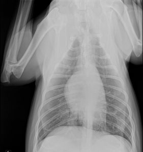
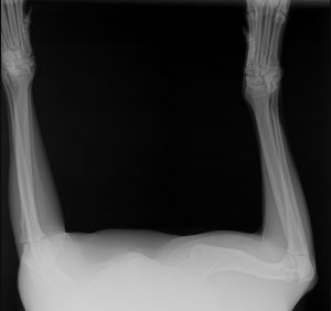
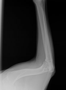
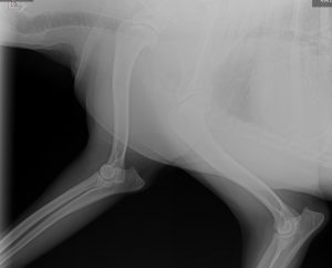
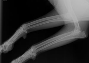
Diagnostic results: Radiographs of bilateral front limbs did not reveal any orthopedic abnormalities (Photo 2-6).
Problem List
1- Warm, firm, painful left triceps brachii muscles
2- Decreased range of motion in left elbow, with repeatable pain response on extension
3- Multiple taut muscle bands in the cervicothoracic region bilaterally (sternocephalicus muscle, cleidocephalicus muscle and trapezius muscle)
4- Left side, latissimus dorsi tender/reactive to palpation along its entire length
5- Taut superficial pectoral at insertion
Differential Diagnoses for Top Two Problems
-Vascular: Blood clots and deficits in micro-circulation can cause local ischemia and subsequent pain.
-Infectious: Lyme or Ehrlicia can cause systemic infection and can also affect the joints, resulting in lameness
-Neoplastic: Osteosarcoma can cause severe pain and lameness with associated bone lesions. The proximal radius/ulna are common locations for osteosarcoma.
-Degenerative: Degenerative joint disease-causing osteoarthritis and possible osteochondrosis dissecans, chronic degenerative intervertebral disc disease.
-Iatrogenic: Insertion of intra-venous catheters can cause vasculitis and subsequent pain
-Congenital: Elbow dysplasia and/or osteochondrosis dissecans
-Auto-immune: Diseases such as lupus or pemphigus affecting the foot-pads, severe allergies causing traumatic chewing/licking, or immune mediated poly-arthritis can lead to pain/lameness
-Traumatic: Soft tissue sprain, strain, bursitis, bruise, hairline fracture(s) or acute intervertebral disc disease.
-Endocrine/metabolic: Obesity can add extra weight on joints
-Myofascia: Soft tissue structures surrounding an injury act in a compensatory manor to guard the area of injury leading to myofascial inflammation and restriction from a sub clinically or clinically altered gait.
Putative/Definitive Diagnosis
I believe that traumatic injury to the triceps muscles led to Cato’s acute left front lameness. Soft tissue injuries typically take months of rehabilitation to fully heal, therefore the re-occurring left front lameness is likely a flare of the same injury. I believe the remainder of abnormalities appreciated during the myofascial examination are all secondary and compensatory to the original traumatic injury. Radiographs ruled out any orthopedic abnormalities including elbow dysplasia, osteoarthritis, osteochondrosis dissecans lesions or lytic lesions indicative of bone neoplasia. Cato receives monthly flea/tick preventative, therefore tick-borne diseases such as Lyme or Ehrlicia are less likely. Daily exercise and appropriate diet improved Cato’s weight and he lost about 5 pounds over 18 months, therefore obesity is not a cause of Cato’s lameness. Cato does not show signs of systemic disease, he does not have any dermatologic lesions and does not exhibit shifting leg lameness, therefore auto-immune causes are not likely.
Medical Decision Making
Cato received one month of weekly treatment, one month of bi-weekly treatments and monthly maintenance treatments. Cervical spine, proximal forelimb and thoraco-lumbar region were targeted during treatments. Cato’s treatments consisted of a combination of dry needling and massage. Cato thoroughly enjoyed his dry needling and a marked parasympathetic response was seen during all sessions.
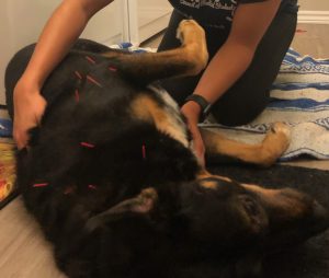


Medical Acupuncture and Related Techniques
Seirin J-type coated needles were used for each treatment. No.1(0.16)x15mm needles were used for points on the distal limb and No.1(0.16)x30mm needles were used for all other points. Sessions began with insertion of GV 20 and Bai Hui. Cato was then asked to lay in either right or left lateral. Points were inserted on either the left or right side, left in for 20 minutes and needles were removed. Cato was asked to lay on the opposite side and process was repeated. Total treatment lengths were typically between 40-45 minutes.
Dry needling points were chosen in order to provide neuro-modulation to all aspects of the nervous system and the myofascia.
Multiple points along the Gallbladder and Bladder channels stimulated the Central Nervous System and were also used regionally for trigger points and pain. The Autonomic Nervous System was specifically targeted using LU 7, LI 4, GV 14, GV 20 and Bai Hui. The peripheral nervous system and myofascial planes were targeted as outlined below:
-Cervicothoracic region: BL 10, BL 11, BL 13, BL 14, BL 15, BL 17, GB 20, GB 21, GV 14, GV 20, cervical spinal nerve points along the course of the omotransversarius muscle
-Shoulder: LU 1, SI 9, SI 12
-Elbow: LU 5, LI 10, LI 11, LI 15, SI 11, TH 10
-Thoracolumbar region: BL 18, BL 19, BL 20
Outcomes, Discussion
Over Cato’s treatments, there was a noticeable decrease in the occurrence of visible lameness in the left front forelimb, and his taut bands and trigger points in the cervical spine improved. Cato seemed to enjoy his treatments, as he often fell asleep while the needles were left in for 20 minutes, and after the needles were removed, he would stand and stretch. I believe that maintenance acupuncture treatments will help decrease chronic pain associated with Cato’s soft- tissue injury, as well as reduce local inflammation during flares of his original injury sustained in July 2018. I believe that Cato would be an excellent candidate for electro-acupuncture and photomedicine when machines are available for use. Cato’s series of treatments shows subjective evidence that acupuncture can provide adequate analgesic properties in the absence of an oral non-steroidal anti-inflammatory medication, as all anti-inflammatories were discontinued following his severe gastric ulceration. Therefore, acupuncture may be an adequate mode of analgesia for animals that do not tolerate non-steroidal anti-inflammatory medications.
References
1. Muscle disorders and rehabilitation in canine athletes. Steiss JE. Veterinary Clinics of North America: Small Animal Practice. 2002 Jan;32(1):267-85.
2. Ultrasonographic and anatomic study of the canine elbow joint. Villamonte-Chevalier AA., et al. Veterinary Surgery. 2014 Jul;44(4):485-493.