Written by a CuraCore Veterinary Medical Acupuncture course graduate. Signed release obtained from client/author. 10S2018046
Abstract
Charlotte, an 11 year-old, spayed female German Shepherd dog presented with a chronic history osteoarthritis and pain in multiple joints and an ongoing lameness in the left forelimb and right hind limb. After minimal improvement through medical management along with veterinary spinal manipulation therapy, her owners sought out integrative and rehabilitative services to help manage Charlotte’s discomfort and improve her lameness. Charlotte received nine treatments using acupuncture, photobiomodulation, pulsed electromagnetic field therapy and massage, resulting in improvements in her lameness score, percent weight-bearing and overall quality of life.
Present Illness/Chief Complaint:
Charlotte is an 11 year old, spayed female, German Shepherd dog that presented for continued arthritic pain and lameness most evident in the right hind limb and left forelimb. Her history includes a right tibial plateau leveling osteotomy (TPLO) for a cranial cruciate ligament (CCL) tear in 2011. A radiographic report from November of 2017 indicated that Charlotte had severe osteoarthritis (OA) in the right stifle, moderate OA in the left stifle, mild OA in the right elbow and severe OA in the left elbow. In March of 2018 she was diagnosed with bilateral carpal hyperextension, with the right being more affected than the left. She currently wears a soft carpal support on the right.
Charlotte has difficulty on slippery and uneven surfaces, long walks, stairs, rising from a sit and running, and currently limps while walking. Her clinical signs are prevalent throughout the day, but appear to be the worst in the morning, after activity and following periods of rest.
Current treatment for Charlotte’s arthritic pain include: Carprofen (1 75mg tab PO BID), Tramadol (1 50mg tab PO BID), Cosequin DS (1 tab PO BID) and monthly veterinary spinal manipulation therapy (VSMT).
PE and Assessments:
O: Charlotte’s weight was 71.6 lbs, with a body condition score (BCS) of 5/9. Her CSU chronic pain score was a 2/4. She was bright, alert and responsive (BAR). Her heart rate was 132 bpm. Cardiopulmonary auscultation was within normal limits (WNL) with no audible murmurs, crackles or wheezes. Lymph nodes palpated soft, smooth and symmetrical. Abdominal palpation was soft and nonpainful. Pupillary light reflex (PLR) was four seconds in both eyes.
Gait: Charlotte was lame on all 4 limbs, with the most significant issues being the left forelimb (L FL), grade 4/5, and a right hind limb (R HL) lameness, grade 3/5. A moderate head bob was noted with L FL paw placement. Her HL gait was very stiff with minimal flexion of her hocks and stifles bilaterally. Charlotte’s FL gait was short and choppy. Initial Examination .MOV
Posture: Charlotte stands with a predominantly level back. Her FLs and HLs are narrow-based. She can sit squarely. She cannot lay in a sternal position, preferring to rest onto a hip. Her transitions required moderate to significant effort. Volitional neck range of motion (ROM) was good overall. Her spinal flexion is limited to her rib cage on both sides.
Neurologic Evaluation: Hopping was absent in her L HL today, but normal in all other limbs. Conscious proprioception (CP)/postural reaction and withdrawal was normal in all limbs. Sciatic, patellar and cranial tibial reflexes were normal in both HLs. Biceps and triceps reflexes were normal in both FLs. Charlotte responded appropriately to all cranial nerve tested (bilateral menace, PLR, facial sensation, and blinking).
Orthopedic Evaluation: Left hock – decreased flexion noted. Left stifle – mild crepitus and decreased extension noted with bony thickening. Right stifle – severe crepitus, decreased flexion and decreased extension was noted with bony thickening. Hips – decreased extension (R>L) with associated discomfort and decreased abduction noted bilaterally. Mild to moderate crepitus was noted in the R hip. Left carpus – moderate crepitus, decreased flexion and mild to moderate carpal hyperextension. Right carpus – increased anterior/posterior (AP) laxity, decreased flexion with associated discomfort, and moderate to severe carpal hyperextension. Left elbow – mild crepitus, decreased flexion, and extension noted with thickening over both medial and lateral aspects. Right elbow – decreased flexion and extension noted with bony thickening more prominent over the lateral aspect. Right shoulder – decreased extension noted. Left HL toes – decreased extension noted at the level of the P1-P2 of digits 3 and 5. Left FL toes – decreased extension and thickening of the P1-P2 joint noted on digit 5. R FL Toes – intermittent crepitus noted at the P1-P2 joint of digits 2 and 3. Right HL toes palpated normally today. Additionally crepitus was palpable over the lumbar spine during movement.
Myofascial/Muscular Evaluation: Moderate myofascial back pain was noted from T6-L2 and severe myofascial back pain was noted from L2-S1. Muscle atrophy was noted over the following regions: thigh atrophy = moderate; shoulder atrophy = mild to moderate; paraspinal muscles = moderate. The following muscles were tense on palpation: both biceps brachii, both pectorals, both sartorius, L tensor fascia latae (TFL), L semimembranosus, and both iliopsoas (R>L).
Problem List
Charlotte’s problems include a grade 4/5 L FL lameness, grade 3/5 R HL lameness, evidence of degenerative joint disease (DJD) in multiple joints (both carpal joints, both elbows, R shoulder, both stifles, L hock, both hips, lumbar spine and toes), diffuse muscle atrophy, bilateral carpal hyperextension, myofascial back pain from T6-S1 and several tense muscles.
Differential Diagnoses
1. Lameness
Vascular – FCE
Infectious – lyme disease, fungal osteomyelitis
Neoplastic – osteosarcoma
Degenerative – multifocal DJD, spinal stenosis, hip dysplasia
Iatrogenic/Intoxication – overuse, result of chronic compensatory gait changes due to pain
Congenital – elbow dysplasia, hip dysplasia
Autoimmune – immune-mediated polyarthritis
Traumatic – undiagnosed fracture, implant failure from previous TPLO
Endocrine/Metabolic – hyperadrenocorticism, hypothyroidism
2. Diffuse muscle atrophy
Vascular – FCE
Infectious – toxoplasmosis
Neoplastic – as part of paraneoplastic syndrome secondary to lymphoma or an insulinoma
Degenerative – sarcopenia due to aging process vs. disuse atrophy from chronic DJD
Iatrogenic/Intoxication – decreased appetite due to tramadol
Congenital – muscular dystrophy
Autoimmune – polymyositis
Traumatic – unlikely
Endocrine/Metabolic – hypothyroidism, chronic kidney disease, nutritional imbalance
Definitive Diagnoses
Based on reported radiographic findings, goniometry and palpation DJD is Charlotte’s putative diagnosis, with evidence of DJD in both carpal joints, both elbows, the right shoulder, both stifles, left hock, both hips, lumbar spine and toes). Recent bloodwork showed no signs of active infection, inflammation or thyroid disease. Although immune-mediated causes cannot be ruled out without the performance of a joint tap, the lack of swelling and the relatively low level of pain described by the owners and elicited on exam make this diagnosis less likely. Additionally, the radiograph report indicated no evidence of fractures or tumors. I attribute the muscle tension and generalized atrophy to disuse and compensatory gait patterns due to chronic pain from ongoing DJD.
Medical Decision Making
Given Charlotte’s presentation, the chronicity of her problems and the number of joints and muscles it was elected to bring Charlotte in for once weekly “integrative” sessions that would include acupuncture, massage, LASER and pulsed electromagnetic field (PEMF) therapy. Each week she was evaluated from a myofascial standpoint so that her massage and other modalities could be tailored for her treatment. Tense muscles were predominantly targeted with massage and LASER therapy. While dry needling acupuncture points were chosen to help with the arthritic and some compensatory back pain through neuromodulation, with particular emphasis place on the right coxofemoral joint, right stifle, both elbows, and associated spinal cord segments.
Acupuncture and Related Techniques
Charlotte’s treatment consisted of nine treatments. Initially the appointments were scheduled on a weekly basis, until her formal recheck at one month. Based on her improvements and the owner’s wishes, it was recommended that her appointments be spaced out to every other week.
Dry needle acupuncture was performed using 0.2x30mm J-type coated Seirin needles. Needles remained in place for approximately 20 minutes each session. An increase in the number of points used occurred over three visits, eventually including all of the following points as tolerated (some minor variation did occur during visits):
– Autonomic/central nervous system: ST 36, BL 13, BL 21, BL 23, BL 25, BL 27, BL 28 (all treated bilaterally), GV14, Bai Hui
– Peripheral nervous system:
o Elbows – LI 11, LI 10, LU 5, PC 3, HT 3 (bilaterally as tolerated)
o Stifles – ST 36 (bilaterally)
o Hips – GB 29 (L), GB 30 (L), BL 54 (L)
o Hocks – KI 3/BL 60 (bilaterally)
Upon Charlotte’s third visit I began implementing electroacupuncture using an Ito ES-130 portable unit dialed to a comfortable frequency, typically around 2 Hz. The leads were placed from BL 28 to BL 21 x2 and ST 36-L to GB 30-L to further augment her treatments for her myofascial back pain and L stifle discomfort.
A class IV Companion 12W LASER using the following presets with a continuous wave: 60-80lb, long coat, light skin, medium coat, max OA. Treatment protocols for the affected joints and surrounding muscles are listed below. The thoracic and lumbar spine were eventually dropped, as more massage was incorporated into Charlotte’s sessions.
– Carpus (bilaterally): 3:09 minutes, 1323 Joules (J), 7 watts (W)
– Elbows (bilaterally): 4:27 minutes, 2403 J, 9W
– Stifles (bilaterally): 5:36 minutes, 3360 J, 10W
– Thoracic spine: 2:30 minutes, 1500 J, 10W
– Lumbar spine: 5:02 minutes, 3020 J, 10W
Effleurage and petrissage massage was performed on the following regions prior to needle placement and while the needles were in place: thoracic and lumbar paraspinals, pectorals, shoulders, inner and outer thighs. Manual therapy locations chosen based on areas of restriction and tension.
Pulsed electromagnetic field (PEMF) therapy was applied to the entire body using the Respond Systems therapy mat for its anti-inflammatory and pain relieving effects.
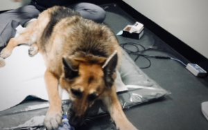
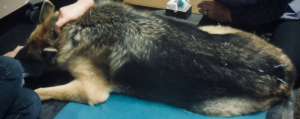
Before Session 4 After Session 4
Outcomes, Insights and Discussion
From Charlotte’s initial visit to her 1 month recheck, Charlotte’s owners reported that Charlotte was moving better and overall seemed more comfortable, particularly the first few days following her appointments. During this time there were improvements in her R HL lameness (grade 2/5) and L FL lameness (grade 3/5). She maintained muscle mass in the thighs and became more symmetrical in the shoulders. The percent weightbearing also showed mild improvement on the R HL using the Companion Stance Analyzer. Although Charlotte’s measurable outcomes only showed mild improvements, I am pleased with these results given the chronicity and the disease process of DJD. After Session 4.MOV
I do not believe one single modality made the difference in Charlotte, but rather a multimodal approach. The use of the LASER, acupuncture, PEMF and massage allowed us to treat Charlotte based on her needs and in entirety. Part of the way through her treatment it was discovered that she responded better with massage rather than LASER therapy to her tense epaxials. It was at this point we altered her LASER protocol and allowed more time for more massage. This reminded and reinforced the need to always reassess your treatment. What works well for one patient, may not be appropriate or as useful in the next patient.
References:
1. Lane DM, Hill SA. Effectiveness of combined acupuncture and manual therapy relative to no treatment for canine musculoskeletal pain. Can Vet J. 2016;57(4):407–414.
2. Dommerholt, JanChou, Li-WeiFinnegan, MichelleHooks, Todd et al. A critical overview of the current myofascial pain literature. Journal of Bodywork and Movement Therapies. 2019 Apr; 23 (2):295 – 30