Written by a CuraCore Veterinary Medical Acupuncture course graduate. Signed release obtained from client/author. 10S2019005
Abstract
This case report describes the integrated treatment and progress of a dog who presented acutely lame on his right pelvic limb. This report also discusses the muscle atrophy and neurologic abnormalities resulting in myofascial changes and pain. The dog’s treatment protocol consisted of 4-weekly sessions involving acupuncture, massage, electrical stimulation, and rehabilitation exercises.
History
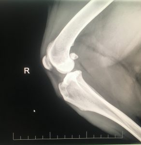
Boba Heath is an alert, energetic, 65-pound (BCS 6/9), 11 year-old, male neutered, Black Mouth Cur who was adopted as a puppy and has a previous medical history of otitis externa bi-annually. Boba lives with a family of four, including two young, active children. His home consists of 80% hard wood flooring and 20% carpeting. His daily routine includes several stairs to outside and stairs to his food and water. Boba’s main exercise is running the backyard. He presented for an acute lameness of his right pelvic limb after slipping on the hard wood floor and falling down. His attitude, appetite, urination and defecation were normal on presentation. Boba does not have a history of gastrointestinal disease. He had mild to moderate dental calculus and severe ceruminous discharge and swelling of his right ear. Boba presented with a grade 4/5 lameness of his right pelvic limb with effusion and medial buttress of the stifle joint. He had positive thrust and drawer of his right stifle, with normal range of motion of his right hip and tarsus. Stifle radiographs revealed effusion and osteophytes in his right stifle joint (photos #1 and #2). A tentative diagnosis of a partial cranial cruciate ligament tear and otitis externa was made. Boba was sent home with Posatex Otic and Galliprant 60mg PO daily. The client was instructed to limit Boba’s exercise to leash walks for elimination purposes and to avoid explosive running after squirrels, and to return for a re-evaluation of his stifle in two weeks. The goals of therapy included resolution of his otitis externa and improvement of Boba’s right rear leg lameness.
Physical Exam
During Boba’s follow-up examination, his owners reported his lameness had improved, but not resolved over the previous two weeks. Boba has had no change in his bark or exercise intolerance. His owners have not heard him clear his throat. A gait analysis revealed a 3/5 grade lameness of his right pelvic limb with a reluctance to circle to the right (videos #1 and #2), and a propensity to lean to the left while walking. A neurological exam indicated bilateral normal conscious proprioception reflexes, hypo-reflexive patellar reflex on the right, and a normal patellar reflex on the left. Boba’s panniculus reflex was normal on the left from his tail base cranially to his thorax, but absent from his tail base extending cranially to the 5th lumbar vertebrae (L5) level on his right side. Withdrawal of both hind limbs was normal and he did not have a crossed extensor reflex on either hind limb. On myofascial exam, kyphosis was noted from his 1st lumbar vertebrae (L1) extending caudally to L5, bilateral tenderness to palpation was found along his longissimus dorsi from L1 to L5, taut bands bilaterally in his trapezius muscle, and atrophy of his sartorius muscle with a subjective hypertrophy of the biceps femoris. Boba turned to look and licked his lips during gentle palpation of his lumbar spine; he did not react with manipulation of either stifle. His effusion was resolved in his right stifle, and no medial buttress was palpable. His stifle was stable upon thrust and drawer manipulations, however, consistent with his radiological indication of osteophytes; his joint was thickened medially and laterally. His otitis externa was resolved.
Problem List
1. Grade 3/5 lameness of the right pelvic limb
2. Hypo-reflexive patellar reflex on the right
3. Absent panniculus from his tail base to L5
4. Kyphosis from L1 to L5
5. Discomfort of his lumbar musculature, specifically L1 to L5 extending bilaterally to the inner Bladder line.
6. Muscle atrophy of his sartorius muscle, possible resultant hypertrophy of his biceps femoris
7. Taut bands bilaterally of his trapezius muscle
8. Otitis externa, resolved at this time
Differential Diagnoses
Right pelvic limb lameness
Vascular: vasculitis, aortic thromboembolism.
Infectious/Inflammatory: foreign body abscess, coccidiodomycosis, infectious synovitis, tick borne illness.
Neoplastic: hypertrophic osteopathy, synovial sarcoma, osteosarcoma.
Degenerative: osteoarthritis, intervertebral disk disease, degenerative joint disease.
Iatrogenic/Intoxication: ingested toxin, which resulted in damage to the cartilage and/or the spinal column.
Congenital: degenerative joint disease, hip dysplasia, osteochondritis dissecans.
Autoimmune: polymyositis, polyneuritis, polyarthritis.
Traumatic: cranial cruciate ligament rupture, iliopsoas muscle strain, hip luxation, stifle luxation.
Endocrine/Metabolic: inadequate nutritional intake resulting in bone, cartilage, or synovial fluid changes.
Myofascial: trigger points in his lumbar musculature, myofascial pain syndrome, atrophy of his sartorius.
Hypo-reflexive patellar reflex and absent panniculus
Vascular: fibrocartilaginous emboli, stroke, embolus.
Infectious/Inflammatory: granulomatous meningoencephalitis, spondylitis of the lumbar spine
Neoplastic: lymphoma of the spinal cord, multiple myeloma of the vertebrae.
Degenerative: intervertebral disk disease, degenerative joint disease, spondylosis.
Iatrogenic/Intoxication: ingested toxin, which resulted in damage to spinal column and/or associated nerve branches in the lumbar spine.
Congenital: vertebral malformation.
Autoimmune: immune-mediated arthropathy, polyneuritis, Geriatric Onset Laryngeal Paralysis and Polyneuropathy.
Traumatic: intervertebral disk disease.
Endocrine/Metabolic: inadequate nutritional intake resulting in abnormal neurological development.
Myofascial: decreased patellar reflex secondary to myofascial pain and restriction of the stifle, absent panniculus may be normal for this patient.
Putative Diagnosis:
A tentative diagnosis of degenerative intervertebral disk disease of the lumbar spine (L4-L5 region), as well as a secondary acute on chronic flare of his right stifle osteoarthritis and partial cruciate ligament injury (CCL). Abnormal neurological signs, including unilateral loss of panniculus and a delayed patellar reflex could be consistent with a chronic thoraco-lumbar disc disorder. It is possible Boba injured his cruciate ligament when he slipped and fell on the hardwood floor. His myofascial changes, including the taut bands in his trapezius muscle, back pain and kyphosis, reflect off-weighting and weight shifting from his hind to his front end.
Medical Decision Making and Medial Acupuncture and Related Techniques Used:
I discussed with Boba’s owner additional imaging and/or referral to a neurologist to diagnosis his specific lumbar spine affliction. The owners declined and elected to pursue acupuncture and related techniques.
Boba’s treatment protocol was designed to address the central and autonomic nervous system as well as peripheral points. Points used are listed in order of placement for all therapy sessions, and if paired points, needles were placed bilaterally unless otherwise indicated.

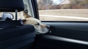
First therapy (10/26/19): For his initial acupuncture treatment, 0.16 x 30mm Seirin needles were placed in all the following points. GV14 (autonomic) and GV20 (autonomic), Bai Hui (autonomic, central input), BL15 (central input, area of taut bands which relaxed soon after needle placement), BL25 (central input, myofascial), BL27 (central input, myofascial), ST36 (autonomic, peripheral input), GB29 (peripheral input), BL23 (central input, myofascial). Needles were in place for 15 minutes. Boba was initially anxious, but calmed and laid down during treatment (photo #3). After the needles were removed, I massaged Boba along his trunk with effleurage and skin rolling techniques for 5 minutes. Boba’s kyphosis resolved post treatment and he was relaxed. Photo #4 was taken by the owner on their way home after therapy.
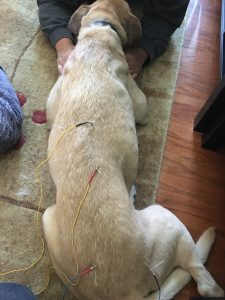
Second therapy (11/2/19): His lameness was still a grade 3/5. Kyphosis was present from L1-L5, with trigger points at BL14, but no taut bands in his trapezius. 0.22 x 25mm Carbo needles were used in all points except as listed. GV14 (autonomic), Bai Hui (autonomic, central input), BL21 (central input, myofascial), BL23 (central input, myofascial), BL24 (central input, myofascial), GB30 (peripheral input, myofascial), ST36 right side only (autonomic, peripheral input, myofascial), ST34 right side only (peripheral input, myofascial), BL14 (central input, myofascial). 0.16 x 15mm Seirin needles were placed in ST36 and ST34 due to tenderness. Needles were in place for 20 minutes. Electrical stimulation (Pantheon manufacturer) for 10 minutes of dense dispersed therapy (2Hz and100Hz) was utilized from BL21 – BL24 and GB30 – Bai Hui (photo #5). Similar resolution of his kyphosis and trigger points occurred post treatment. Boba was relaxed and happy but did not tolerate massage at the end of this therapy session.
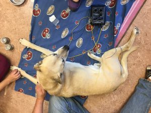

Third therapy (11/9/19): His lameness improved to a grade 2/5. Boba’s owner reported he is moving quicker and more consistently using his right pelvic limb. His myofascial exam indicated tension and tenderness of his iliocostalis from T13 to L5, along the inner Bladder line. He also had tenderness on the lateral aspect of his right stifle joint. His lumbar spine was not kyphotic at the time. 0.22 x 25mm Carbo needles were used. GV14 (autonomic), Bai Hui (autonomic, central input), BL21 (central input, myofascial), BL25 (central input, myofascial), ST36 right side only (autonomic, peripheral input, myofascial), ST34 right side only (peripheral input, myofascial), GB30 (peripheral input, myofascial). Needles were in place for 20 minutes. Electrical stimulation (Pantheon manufacturer) for 10 minutes of dense dispersed therapy (2Hz and 100Hz) was utilized from ST34 to GB30 on his right side and BL21 to Bai Hui on his left side (photos #6 and #7). Boba’s iliocostalis tenderness was resolved post therapy and he allowed massage therapy (effleurage and skin rolling) of his lumbar spine for 5 minutes.
Video #3, 11-16-19, Boba slow walk, post 4th treatment Video #4, 11-16-19, Boba post 4th treatment Video #5, 11-16-19, Boba fast walk, post 4th treatment
Fourth therapy (11/16/19): Boba’s owner reported his lameness is the same as the previous week; however, Boba has been more active and has resumed jumping up on his pelvic limbs again. In the clinic, his lameness was a grade 2/5. On myofascial exam, he had kyphosis from L1-L5, as well as tension and tenderness of all peripheral points needled. 0.22 x 25mm Carbo needles were used. GV14 (autonomic), Bai Hui (autonomic, central input), BL21 (central input, myofascial), BL25 (central input, myofascial), ST36 (autonomic, peripheral input, myofascial), ST34 right side only (peripheral input, myofascial), GB29 (peripheral, myofascial), BL40 right side only (peripheral, myofascial). No electrical stimulation was utilized. Needles were in place for 20 minutes followed by massage (effleurage and skin rolling) of his lumbar spine for 10 minutes. Post treatment videos #3 and #5 demonstrate the speed dependent difference his in his gait, while post treatment video #4 demonstrates Boba walking away and toward the camera.
Discussion:
After Boba’s third therapy, I consulted with a certified rehabilitation therapist. After evaluating Boba’s videos, the therapist recommended home exercises of sit to stands (10 repetitions BID), front-ups on stairs or a box (10 repetitions BID), and for the owner to encourage Boba to stand on his hind legs for 10-20 seconds at a time BID. Boba’s treatment has continued, both at home and in the clinic, after his fourth therapy. He is still a grade 2/5 lameness at a walk, but when he runs and plays, his lameness is resolved. His mobility has improved both with Galliprant administered at home and due his acupuncture treatments stimulating his nervous system, both centrally and peripherally, and addressing his myofascial pain. A notable improvement was seen with the addition of at-home physical therapy.
Considering the possibility of a CCL injury, his improvement with anti-inflammatories and rest was quicker than expected. It is possible Boba does have a chronic CCL injury; however, it may manifest itself as periodic lameness and inflammation of his knee or if his lameness is a result of referred pain. His atrophy of his sartorius has improved from analgesics, nerve stimulation by electro-acupuncture, and from the at-home physical therapy exercises. Due to the concern of both intervertebral disk disease and recurring knee inflammation, it is recommended Boba continue acupuncture and related techniques for the rest of his life. In addition to addressing Boba’s musculoskeletal issues, additional needle points will be added for his recurring otitis externa. Boba continues to be happy and playful.
Pelvic limb lameness in dogs is a common complaint in general practice and is often related to cranial cruciate ligament injury. Boba’s systemic physical, neurological, and myofascial exam highlight the underlying and/or complicating factors contributing to a pelvic limb lameness and possible intervertebral, degenerative pathology. The need to assess the entire patient, at every appointment, is evident in this case.
I found no published scientific references for the use of acupuncture and physical therapy as a management strategy for cranial cruciate ligament disease in the dog, but there are several journal articles regarding acupuncture and related techniques for knee injuries in humans. One article, listed below, did include the use of acupuncture post CCL surgical repair in the dog. Several conference proceedings discussed use of integrative medical management for cruciate ligament injuries in the dog. The use of integrative medical management for the management of cranial cruciate ligament disease is a much needed area of study in the veterinary field.
I learned the importance of assessing the entire patient, at every appointment. It is also concerning how many of my previous patients with CCL injury may have undergone surgery with underlying, complicating issues, and who may have been better served with integrative medical management. I have educated my colleagues regarding the importance of a neurological and myofascial exam for every patient, especially those presenting with a lameness.
What is also striking about this case is the increased bond created between myself, Boba, and my clients during his treatment course. Boba is always happy to see me and becomes very excited when he knows it is time for his therapy. I enjoy watching him progress through his recovery.
References:
Shmalberg DVM, DACVN, Justin. “A Randomized Controlled Blinded Clinical Trial of Electro-acupuncture Administered One Month After Cranial Cruciate Ligament Repair in Dogs”. Jessica Burgess DVM, Wendy Davies CVT. American Journal of Traditional Chinese Veterinary Medicine, Vol 9, No.2, August 2014.
Li X. “Acupuncture for Myofascial Pain Syndrome: A Network Meta-Analysis of 33 Randomized Controlled Trials”. Wang R, Xing X, Shi X, Tian J, Zhang J, Ge L, Zhang J, Li L, Yang K. Pain Physician. 2017 Sep; 20(6):E883-E902.
Conference Proceedings:
Robinson, Narda G., “Non-surgical, Integrative Medical Approaches to Cranial Cruciate Ligament Disease (CrCLD) in Dogs” Conference Proceedings: New York Vet Show: NY Vet Show 2018.
Todd, Gregory, “A Traditional Chinese Veterinary Medical Approach to Musculoskeletal Diseases” Conference Proceedings: Wild West Veterinary Conference: Wild West Veterinary Conference 2013.
Todd, Gregory, “The Treatment of Musculoskeletal Disease Utilizing Traditional Chinese Veterinary Medicine” Conference Proceedings: Wild West Veterinary Conference: Wild West Veterinary Conference 2016.