Written by a CuraCore Veterinary Medical Acupuncture course graduate. Signed release obtained from client/author. 10L2019009
Abstract:
A middle-aged gelding with chronic (>one year) mild hind and front limb gait asymmetry was treated with a combination of electroacupuncture, acupuncture, and laser on four separate occasions over one month. A noticeable improvement in gait quality was noted following the third treatment and a change in demeanor was reported by caretakers following the second, third, and fourth treatments.
History and Presentation:
The 14-year-old warmblood gelding named “Berkley”, was examined on December 10, 2019 for a re-evaluation of an ongoing fore and hindlimb gait asymmetry. Berkeley had previously been diagnosed with mild bilateral stifle instability, a left hind lateral suspensory branch desmitis, mild left forelimb pastern and coffin joint degenerative joint disease, and moderate left and right carpal degenerative joint disease.
The horse was treated with shockwave therapy multiple times for the left hind lateral distal suspensory desmitis, but continued to have an asymmetric hind and fore limb gait despite improvement of the lesion on ultrasound and resolution of the original clinical signs (heat, pain on palpation, and a decreased ability to fully perform his required work). An intensive rehabilitation-based exercise protocol had been prescribed and followed throughout his convalescence to attempt to build hind end musculature to stabilize the stifles and prevent fatigue related suspensory strain.
Berkley Dec 10 2019 Away Video Berkley Dec 10 2019 toward video Berkley Dec 10 lateral Video
Physical Examination/ Clinical Assessments:
First General Physical Exam (10/12/2019)
The ongoing asymmetry of this gelding’s hind end could be described as a hyperextension of the left hind fetlock at the weight bearing phase of the trot and an intermittent short stride of the right hind limb at the trot. Proximal flexion tests of the left and right hind limbs resulted in a shortened stride for approximately 15m post flexion at the trot for each limb. Distal hindlimb flexion tests resulted in a slightly longer abnormal gait for the left hind than the right hind. The front-end asymmetry was only visible for the first couple of circles on the lunge line traveling in a counter- clockwise direction. Proximal flexion tests of the front limbs were both positive with the left fore showing a prolonged shortened stride post flexion than the right.
Myofascial Palpation Evaluation (10/12/2019) before first session:
Diagnostic Acupuncture Exam “DAPE” (specific reactive points):
Berkeley was reactive to even light touch over ST 10 on the right side and LI 18 on the right side.
Myofascial Exam:
There was marked myofascial restriction palpable over the cranial and caudal gluteal muscles on the left side. A decreased range of motion of the sacroiliac joint and lumbosacral region was noted on the right and left side. Muscle hypertonicity was palpable over the left and right lumbar epaxial musculature but was more pronounced on the left side. A trigger point was discovered immediately cranial to BL 27 on the left side.
Balance:
Feet were replaced evenly and in a normal position.
Cervical Mobility:
Lateral flexion of the second and third cervical vertebrae in both directions was difficult and elicited resentment.
Withers and Scapular Mobility:
No abnormalities noted in these regions.
Shoulder (internal/external rotation):
No abnormalities noted, motion was even on both limbs.
Front limb stretch with neck lowering:
Berkley was able to easily and equally stretch his head towards his front fetlocks when they were held above the ground in front of him.
Joint palpation/Range of Motion:
Both carpi had a decreased range of motion, with the left being more compromised than the right. The left accessory carpal bone had limited motion in a lateral-medial/medial-lateral plane. Moderate intra-articular stifle effusion was palpable on the right, and mild intra-articular stifle effusion was palpable on the left.
Reactivity of paraspinal muscles:
Stimulation of his epaxial musculature from withers to croup elicited a hyperreactive response on the left side, but a normal response on the right.
Lumbosacral spinal mobility:
Motion palpation of the lumbosacral area was mildly resented and decreased bilaterally.
Sacroiliac Mobility and hindlimb circles:
Left side sacroiliac mobility was decreased and circles on this side were more challenging to keep round instead of a diamond shape.
Problems:
Berkley’s problems could be characterized as a mild left hindlimb lameness and a mild left forelimb lameness.
Differential Diagnoses:
1. Mild left hindlimb lameness
Vascular: pain secondary to a thrombus causing vascular compromise.
Infectious: Equine Protozoal Myelitis
Neoplastic: a neoplastic process that compromises the outer aspect of the spinal cord or compresses a nerve outflow tract supplying hind limb musculature.
Degenerative: degenerative joint disease
Iatrogenic/Intoxication: an iatrogenic muscle imbalance due to a poorly designed work regimen or improper shoeing
Congenital: stifle instability due to congenital malformation, progressive distal suspensory desmitis, bone cyst, predisposition to suspensory branch hyperextension and stifle instability secondary to an upright proximal hind limb conformation
Autoimmune: autoimmune myelopathy secondary to herpesvirus
Traumatic: soft tissue or boney lesion from either acute or chronic trauma
Endocrine/Metabolic: a decreased ability to heal soft tissue lesions secondary to Equine Pituitary Pars Intermedia Dysfunction (PPID)
Myofascia: myofascial dysfunction may be a primary cause of hindlimb lameness/ asymmetry, or may be secondary to another underlying cause
2. Mild left forelimb lameness
Vascular: vascular compromise of the navicular bone resulting in bone degenerative disease or vascular compromise secondary to a carpal canal syndrome
Neoplastic: a neoplastic mass not readily found with palpation/observation (keratoma, space occupying mass in and/or around the brachial plexus are two possible examples)
Degenerative: degenerative joint disease of a forelimb joint.
Iatrogenic/Intoxication: mild laminitis secondary to juglone toxicity, improper farriery
Congenital: bone cyst
Autoimmune: autoimmune based myopathy
Traumatic: sole bruising, soft tissue trauma of an acute or chronic nature
Endocrine/Metabolic: a decreased ability to heal soft tissue wear and tear secondary to PPID, or laminitis secondary to PPID
Myofascia: myofascial dysfunction may affect the nerve supply to the front limbs and cause a mild inequity of stride, or may occur as a result of a gait deficit
Definitive/Putative Diagnoses:
1. Mild left hindlimb lameness
Lameness localization with nerve bocks and flexion tests followed by ultrasound exam of the distal suspensory branch and stifle radiographs identified two causes of the hindlimb gait deficit: mild degenerative joint disease likely secondary to a noted mild stifle instability along with a mild chronic suspensory branch desmitis. The conformation of the gelding predisposed him to both hind limb diagnoses, along with his history of schooling and showing at third and fourth level dressage. A normal ACTH result decreased the likelihood of PPID playing a role. The unilaterality of the suspensory lesion does not support a diagnosis of progressive suspensory desmitis, and the history of an acute lameness that responded well to shockwave therapy and following a rehabilitation and return to work plan supported a traumatic lesion of the suspensory. It is hypothesized that the suspensory desmitis may have been from overusing the left hind due to right stifle soreness.
2. Mild left forelimb lameness
Flexion tests, nerve blocks, and radiographs were used to reach a putative diagnosis of minor degenerative joint disease of the coffin and pastern of the left forelimb and moderate degenerative joint disease of the left and right carpus.
Medical Decision Making:
Due to the chronic nature of this gelding’s gait deficits, the goal was to attempt to more fully normalize his gait to achieve a higher level of performance using acupuncture and laser therapy along with his currently prescribed rehabilitation plan. The show schedule planned for the following year made other treatment options such as intra-articular and systemic anti-inflammatory medications difficult.
The initial four treatments were planned to evaluate response and use this information to develop a plan for the 2020 show and clinic season. The favourable response immediately post treatment has led the owner to plan on using acupuncture and laser both for gait improvement but also for the calming effect noted. The once a week schedule used so far worked well for the horse and was financially feasible for the owner, although response suggests that these grouped treatments may not need to be ongoing at that rate and may be decreased to a less frequent basis.

Medical Acupuncture and Photobiomodulation Techniques Used:
December 10, 2019:
Central Nervous System: LI 16 (cervicothoracic spinal nerves) were placed first to gauge the horse’s reaction to needle placement, and as a local point for the neck stiffness and discomfort. Needles were inserted at BL 23 (thoracolumbar spinal nn), BL 26 (caudal lumbar spinal nn), and BL 27 (S1 spinal n) bilaterally as local points for the lumbar epaxial muscle splinting as well as for their effects on cranial gluteal musculature.
Peripheral Nervous System: LI 11 was placed for a distal point for cervical involvement and for immune modulating effects (also ANS point). BL 40 (tibial nerve) was needled bilaterally along with ST 36 (fibular nerve) to neuromodulate the stifle.
Autonomic Nervous System: GV 14 (cervicothoracic spinal nn, also a CNS point) was used for local cervical pain and parasympathetic stimulation. Bai Hui was used for the lumbosacral dysfunction but also to stimulate nerves supplying muscles supporting the stifles. ST 36 (fibular nerve) was chosen for both its PNS and ANS properties.
Myofascial Dysfunction: GB 21 (spinal accessory nerve) was placed to address myofascial tension in the lower cervical area. The most notable areas of myofascial dysfunction were overlapped by the CNS points BL 23, BL 26, and BL 27 that were also used for their CNS effects.
All above points were needled with Seirin J type size number 5 (0.25 x 40 mm) acupuncture needles using the accompanying guide tubes for insertion.
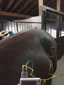
Photobiomodulation was recruited with an Activet Pro LaserShower device with the following wavelength capabilities: laser infrared radiation 950 nm, broadband infrared radiation 850 nm, visible red light radiation 630 nm, and visible blue light radiation 470 nm. This laser was set on 1000 hz and moved slowly for five minutes along the spine from the seventh thoracic vertebrae to the tuber sacrale, and then held over LI 18 (cranial cervical spinal nn) for 6 minutes and on ST 10 (midcervical spinal nn) for three minutes on the right side.
Electroacupuncture was used from BL 23 to BL 26 bilaterally using an ITO set at 1.4 Hz at the “2” frequency for 10 minutes. Berkley was returned to his normal daily turnout schedule following treatment and resumed his regular workout schedule of lunging and riding the following day.
December 17, 2019: Second Physical Exam/ DAPE
Lunging and jogging in hand were not appreciably different from the week before. On palpation there was subtle myofascial restriction over the base of the left neck. The horse responded equally normally to stimulation of the epaxial musculature on the left and the right as opposed to the previous visit. Sacroiliac and lumbosacral mobility was notably improved bilaterally. Trigger points over BL 21 and BL 23 on the left side were noted. Myofascial restriction over left and right coxofemoral joints was noted. Carpal range of motion remained asymmetrical with left still being less than the right.
December 17, 2019: Treatment
The previously described acupuncture points from December 10, 2019 were repeated using the same J-Seirin needles with the following additions:
Myofascial Dysfunction: GB 29 (sacral spinal nn, caudal gluteal n, also CNS effects) and GB 30 (sciatic n, caudal gluteal n) were used to treat the myofascial restriction palpated over the coxofemoral joints. BL21 (thoracolumbar spinal nn) was added for both myofascial dysfunction, but also as a CNS point.
The same ActiVet Pro Laser Shower was used at 1000 Hz along the spine from the seventh thoracic vertebrae to the tuber sacrale for five minutes following by two minutes over both BL 23 and BL 21 on the left to tackle the muscle spasm palpable at that location. The 1000-3000 Hz setting was then used for 10 minutes over the base of the left side of the neck to treat the myofascial dysfunction palpable there.
Electroacupuncture was performed using the Ito to run electricity between BL 23 and BL 26 on both sides for five minutes, and from BL 26-BL 27 on both sides for four minutes on level 3, frequency 2 at the low setting (1.4 Hz).
December 23, 2019: Third Physical Exam/DAPE
Horse was noted to be moving more freely with less warmup time on lunge line. Significant improvement in hind fetlock extension of left hind was noticed and no front-end asymmetry was seen on lunge or when jogged in hand. Physical examination revealed myofascial restriction over the left caudal gluteal muscles. A decreased range of motion of the second and third cervical vertebrae when asked to passively flex neck to the left was noted. A decreased range of motion of the right sacroiliac and lumbosacral areas was also seen in comparison to the left. Carpal range of motion still appreciably decreased.
December 23, 2019: Treatment
The acupuncture protocol described for December 10, 2019 was repeated.
Electroacupuncture was performed again as previously described from BL 23 to BL 27.
Photobiomodulation was recruited using the previously described laser on 1000 Hz for five minutes along the spine from the seventh thoracic vertebra to the tuber sacrale. It was then used over C2-C3 for another five minutes on the same setting to treat the decreased range of motion and discomfort noted there. Finally, it was used over both carpi for ten minutes on each joint.
January 9, 2020: Fourth Physical Exam/DAPE
Berkley was moving very well at all three gaits on the lunge at this follow up examination and in hand at the trot. Owner and rider continued to report an improvement in both rideability and temperament post treatment. Physical examination on this date revealed an ability to flex neck equally in both directions and only a very slight decrease in the left sacroiliac range of motion when compared to the right. Left carpal range of motion remained decreased. A trigger point was present over BL 54 on the left.
January 9, 2020: Treatment
The same previously described J Seirin needles were used as described for the December 10, 2019 treatment with the following additions:
BL 54 (cranial gluteal n, dorsal rami of sacral spinal nn) was needled after the point was lasered for five minutes on the muscle spasm setting to treat the trigger point palpated there. GB 29 (sacral spinal nn, caudal gluteal n, also CNS effects) and GB 30 (sciatic n, caudal gluteal n) were used to treat the myofascial restriction palpated over the coxofemoral joints.
Electroacupuncture was repeated as previously described from BL 25 to BL 27.
Berkley Jan 9 2020 Lateral Video Berkley jan 9 away video Berkley Jan 9 lateral 2 Berkley Jan 9 toward video
Outcomes, Insights, Discussions and References:
The accompanying videos demonstrate the improvement in Berkley’s gait from December 10, 2019 to January 9, 2020. The horse did not receive any other treatment or medication during this period, and his workload has remained consistent throughout the past few months. The multifaceted nature of this horse’s gait deficits coupled with his show schedule that precludes the use of ongoing medication to mitigate those deficits prompted the owner to consider acupuncture to improve his gait. The owner is excited to have this modality to use on an ongoing basis to provide comfort and allow her to carefully maintain and promote musculature to support his stifles and prevent fatigue related fetlock hyperextension. She appreciates the necessity of this to maximize the horse’s potential as he continues to move up the levels of dressage. The idea of using acupuncture to eliminate equine lameness is not new. Studies looking at the use of acupuncture to improve equine lameness are mixed in their results. A controlled clinical trial performed in 1989 (Steiss, et al. 1989) showed no significant differences between control horses and those treated with acupuncture. A study performed in six horses with experimentally induced pain from a Merkens’ method -based shoe that applied pressure to the sole of the horses in the trial was used to cause lameness that affected stride length and character. Acupuncture was found to mitigate but not eliminate the effects of the shoe to a statistically significant degree (Huisheng Xie, Ott, E.A., Colahan, P 2001). More recently “A pilot study of the effects of acupuncture treatment on objective and subjective gait parameters in horses” was performed (Dunkel et al 2017). It concluded that acupuncture was able to effect a positive change in horse gaits in a measurable manner. The study size in all three of these experiments was less than or equal to ten horses and may be responsible for the varying outcomes along with the fact that lameness in many cases may be affected by many other variables such as conformation, other sources and causes of pain, and the possible variability of the acupuncture procedure itself. More work needs to be done to help quantify the case selection for acupuncture in the field of equine sports medicine but positive case results such as this one build confidence in both the horse owner and the veterinarian applying the technique.
Steiss, Janet E, White, NA, Bowen, JM “Electroacupuncture in the treatment of chronic lameness in horses and ponies: a controlled clinical trial.” Can J Vet Res 1989; 53:239-243.
Huisheng, Xie, Ott, E.A., Colahan, P, “Influence of acupuncture on experimental lameness in horses” AAEP Proceedings 2001; 47:347-357.
Dunkel, Bettina, Pfau, T, Fiske-Jackson, A., Veres-Nyeki, K.O., Fairhurst, H., Jackson, K., Chang, Y., Bolt, D.M. “A pilot study of the effects of acupuncture treatment on objective and subjective gait parameters in horses” Vet Anaesth and Analg 2017; 44:154-162.